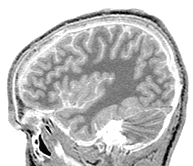
Figure 1: Raw data: sagittal MRI section |

Figure 2: Cerebrum only, cleaned manually. (Orientation and contrast levels have been standardized, so this will not match exactly to the raw original.) |
|
Figure 1: Raw data: sagittal MRI section |
Figure 2: Cerebrum only, cleaned manually. (Orientation and contrast levels have been standardized, so this will not match exactly to the raw original.) |