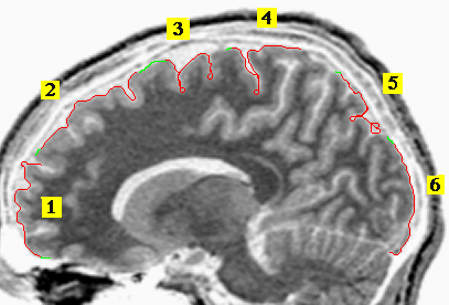
The initial green pixels of trace 3 were used as the training set. The network continued tracing ahead (red pixels) until it ran into trouble, in areas of the contour unrepresented by the training set. The other traces represent restarts of the network-generated tracing (without further training) in new areas of the image (green=manual, red=network). The end of trace 5 shows an area where the brain boundary is indistinct, and confounded by possible ghost structures from the MRI; the network performance is degraded by the region's similarity.